Photoacoustic Mammoscopy for evaluating screening-detected lesions in the breast
Progress
PAMMOTH Results
Summary of the context and overall objectives of the project
With Europe’s aging population, its high incidence of breast cancer, its tightening health-care budget, and room for improvement in conventional breast imaging modalities, there is a need for a technique that can provide images with high specificity, contrast and spatial resolution. Photoacoustic imaging may turn out to be that technique. In photoacoustics, the contrast is dependent on light absorption in tissue. Since blood, fat and water in tissue absorb light depending on the wavelength used, the method can provide spectroscopic (molecular) specificity to these and other tissue constituents. Blood is of interest as a biomarker since more blood vessels are often present at tumor sites.
PAMMOTH brought together experts from various disciplines to work on a new generation system for imaging the breast using both photoacoustics and ultrasound. Academia, industry and the clinic from 6 different countries were part of the PAMMOTH consortium. The consortium’s objective was to develop, validate and begin exploitation of a dedicated breast imaging device. The proposed device would combine non-invasive 3D photoacoustic imaging with ultrasound imaging to provide full-breast, multimodal images to the radiologist. The device has high through-put, possesses no carcinogenic potential, uses no contrast agent and causes no pain or discomfort to patients.
At the conclusion of the project, we have developed the PAMMOTH imager which can extract two imaging contrasts from tissue, and performed first in human studies. From the study on 10 patients patients, the images do not seem as yet to reveal universal and unmistakable PA imaging biomarkers. Yet it has been possible to visualize specific features at tumor locations which may have diagnostic potential. The presence of the ring pattern of photoacoustic intensity around the tumor, and progressive reduction of photoacoustic intensity inwards into the tumor may turn out to be important signatures of breast disease. The contribution of the sound speed has been helpful to provide context to these blood vessel morphologies, as well as identify tumors from healthy surrounding.
Future work will concentrate on continuing the inclusion of patients, and continuing the analysis of the data. The onus will now lie on extracting the information from the PA data which will depend on improved image reconstruction for retrieving the US reflectivity, blood oxygen saturation in and around tumor vessels as well as on improved image processing, image analysis and visualization.
Overview of the results
At the conclusion of the project we have together realized the following:
The industrial partners of the project have made strides in increasing their competitive edge. A new laser product based on the research results was possible. A patent application was also made for a method of depolarization compensation. One of the partners was able to attract a new shareholder. This new investor and further investments from one of the existing shareholders, has enabled to expand the partner's team and continue on the road to bringing 3D PA-US breast Imaging to fruition. The additional investments also serve as matching finances to a number of awarded grant proposals, such as Eurostars 2, EU-REACT, and Dutch SME awards. The academic and hospital partners also have plans for further collaboration.
At this point in we can say that the PAMMOTH imager and the plans that the consortium has to continue collaboration will now make possible to develop a version which lead to a CE-mark where scientific evidence for the efficacy and utility of photoacoustic imaging in various roles can be obtained in well-designed and well-implemented clinical studies. These roles can be in diagnosis; or in monitoring of neoadjuvant chemotherapy; or further in time in screening application can be obtained in well-designed and well-implemented clinical studies.
Progress beyond the state of the art, expected results and potential impacts
Public progress report archive
Please find here a short progress report on the first 18 months of our PAMMOTH project.
Please find here a short progress report on the first 36 months of our PAMMOTH project.
With Europe’s aging population, its high incidence of breast cancer, its tightening health-care budget, and room for improvement in conventional breast imaging modalities, there is a need for a technique that can provide images with high specificity, contrast and spatial resolution. Photoacoustic imaging may turn out to be that technique. In photoacoustics, the contrast is dependent on light absorption in tissue. Since blood, fat and water in tissue absorb light depending on the wavelength used, the method can provide spectroscopic (molecular) specificity to these and other tissue constituents. Blood is of interest as a biomarker since more blood vessels are often present at tumor sites.
PAMMOTH brought together experts from various disciplines to work on a new generation system for imaging the breast using both photoacoustics and ultrasound. Academia, industry and the clinic from 6 different countries were part of the PAMMOTH consortium. The consortium’s objective was to develop, validate and begin exploitation of a dedicated breast imaging device. The proposed device would combine non-invasive 3D photoacoustic imaging with ultrasound imaging to provide full-breast, multimodal images to the radiologist. The device has high through-put, possesses no carcinogenic potential, uses no contrast agent and causes no pain or discomfort to patients.
At the conclusion of the project, we have developed the PAMMOTH imager which can extract two imaging contrasts from tissue, and performed first in human studies. From the study on 10 patients patients, the images do not seem as yet to reveal universal and unmistakable PA imaging biomarkers. Yet it has been possible to visualize specific features at tumor locations which may have diagnostic potential. The presence of the ring pattern of photoacoustic intensity around the tumor, and progressive reduction of photoacoustic intensity inwards into the tumor may turn out to be important signatures of breast disease. The contribution of the sound speed has been helpful to provide context to these blood vessel morphologies, as well as identify tumors from healthy surrounding.
Future work will concentrate on continuing the inclusion of patients, and continuing the analysis of the data. The onus will now lie on extracting the information from the PA data which will depend on improved image reconstruction for retrieving the US reflectivity, blood oxygen saturation in and around tumor vessels as well as on improved image processing, image analysis and visualization.
Overview of the results
At the conclusion of the project we have together realized the following:
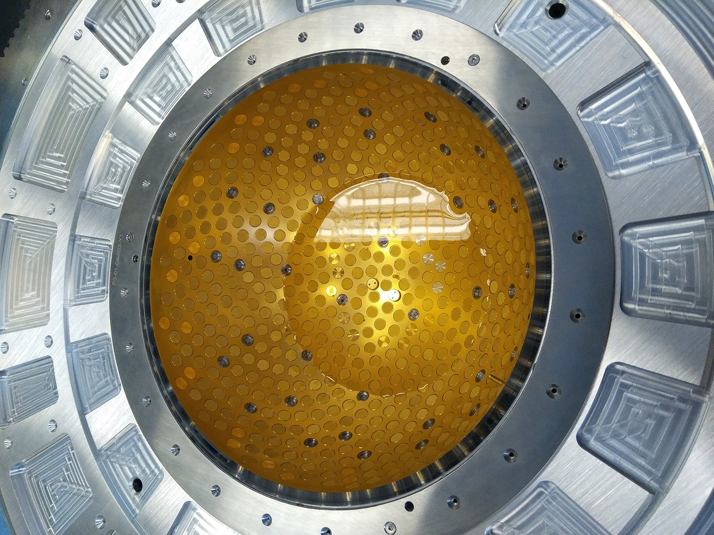
- The clinical prototype PAMMOTH, boasting sub-systems that are technically beyond the state-of-the-art, has been developed, tested and is being used to image patients in a clinical study.
- The developed ns laser has among the highest energy outputs per ns pulse. The OPO pumped by the laser allows fast tuning and high efficiency. The light is coupled to the PAMMOTH imager using an optical fiber bundle, designed to accept high energy pulses, split in 40 outputs.
- The 512 ultrasound transducers arranged in hemispherical geometry provide a low minimum detectable pressure with a wide fractional frequency bandwidth.
- The 512 multi-channel Data-Acquisition System is also equipped to transmit ultrasound from the same transducers for ultrasound imaging.
- Cups of various sizes to immobilize the breast during measurements have been produced to hold the breast in the imaging bowl.
- A novel algorithm has been developed for the specific task of reconstructing intermediate images on the fly as projections are being acquired.
- Novel algorithms for speed of sound reconstruction from ultrasound data have been developed and tested.
- A photoacoustic image reconstruction (using a priori known speed of sound map) based on an iterative approach has been developed.
- The algorithm for quantitative photoacoustic reconstruction has been developed.
- A suite of test and calibration objects has been developed.
The industrial partners of the project have made strides in increasing their competitive edge. A new laser product based on the research results was possible. A patent application was also made for a method of depolarization compensation. One of the partners was able to attract a new shareholder. This new investor and further investments from one of the existing shareholders, has enabled to expand the partner's team and continue on the road to bringing 3D PA-US breast Imaging to fruition. The additional investments also serve as matching finances to a number of awarded grant proposals, such as Eurostars 2, EU-REACT, and Dutch SME awards. The academic and hospital partners also have plans for further collaboration.
At this point in we can say that the PAMMOTH imager and the plans that the consortium has to continue collaboration will now make possible to develop a version which lead to a CE-mark where scientific evidence for the efficacy and utility of photoacoustic imaging in various roles can be obtained in well-designed and well-implemented clinical studies. These roles can be in diagnosis; or in monitoring of neoadjuvant chemotherapy; or further in time in screening application can be obtained in well-designed and well-implemented clinical studies.
Progress beyond the state of the art, expected results and potential impacts
- The combination of photoacoustic and sound speed imaging makes the PAMMOTH imager unique.
- The laser developed produces pulse energies at the fundamental and second-harmonic which are highest within the same application class of lasers.
- The characteristics of the ultrasound detectors are superior to the state-of-the-art detectors.
- A multi-channel Data-Acquisition System (DAS) shows low noise and compared to devices available. The analog front ends in the PAMMOTH DAQ are also capable of driving ultrasound generation by electrical actuation of transducers.
- The 3D phantom developed are among the first for photoacoustics which may be described as semi-anthropomorphic as well as physiopathological phantoms.
Public progress report archive
Please find here a short progress report on the first 18 months of our PAMMOTH project.
Please find here a short progress report on the first 36 months of our PAMMOTH project.